


Surgical treatment of rectal prolapse
Rectal prolapse is the movement of a fragment of the large intestine (rectum) through the anal canal and rectum. There are three main forms of rectal prolapse:
- total prolapse (full wall) - displacement of all layers of the rectal wall through the anus (mucosa, muscles),
- partial prolapse - displacement of a fragment of the rectal mucosa;
- Internal prolapse (hidden, rectal intussusception) - intussusception of one section of the intestine into another, which is not accompanied by the displacement of the rectum outside the anus.
The causes of rectal prolapse are not fully understood.
The causes of prolapse of the rectum in children include chronic constipation and the associated increased effort during defecation. Children suffering from cystic fibrosis are also more susceptible to the disease due to a chronic cough that causes an increase in intra-abdominal pressure.
Rectal mucosal prolapse in adults is often associated with the presence of large rectal varices (hemorrhoids). In the elderly (mainly in older women, who constitute the largest group of patients suffering from rectal prolapse), the factor conducive to the occurrence of the disease may be significant weight loss, which, in combination with weakening of the pelvic and anal muscles, reduces the support for the rectum. In some patients, the presence of symptoms may be related to a history of perinatal trauma.
Chronic constipation and stool output are considered to be the main causes of rectal prolapse in young people. Conditions such as injuries and tumors of the spine and spinal cord, psychiatric disorders, and multiple sclerosis (MS) also contribute to rectal prolapse.
In the initial stage of the disease, prolapse of the rectum is accompanied by defecation. As the discomfort progresses, the symptoms intensify and may accompany situations with increased intra-abdominal pressure, such as sneezing, coughing, and pressure on the stool. In the absence of adequate treatment for the disease, prolapse of the rectum can occur spontaneously, without a cause.
The disease can manifest itself:
- bleeding that is caused by damage to the rectal mucosa
- excretion of increased amounts of mucus,
- constipation,
- incontinence.
The patient may experience pain and more frequent urgency than normal. It is not uncommon for you to feel incomplete bowel movement.
Diagnosis of rectal prolapse:
The basic diagnostic method for rectal prolapse is rectal examination. During the examination, decreased anal sphincter tone is found, while rectal prolapse is observed during pressure on the stool. Rectoscopic examination allows the assessment of about 20 cm of the large intestine using a special sight glass. Rectoscopy performed on a patient suffering from rectal prolapse may show the place of intussusception, the presence of inflammation or damage to the mucosa. Colonoscopy (endoscopic examination of the large intestine) is performed in adults qualified for surgical treatment of rectal prolapse to accurately assess the entire large intestine. In some cases, it is advisable to do so. Hinton test, which allows you to assess the speed of intestinal passage. It involves the oral administration of special markers for several days, which are then visualized in a radiological examination.
Differentiation of rectal prolapse:
Symptoms similar to those seen as a result of rectal prolapse may actually be other disease entities. Rectal prolapse is differentiated from:
- large hemorrhoids (varicose veins),
- rectal tumor moving through the anus,
- genital warts,
- abnormal morphology of the perineum,
- polyps in the anus.
Based on clinical symptoms, rectal prolapse should also be differentiated in inflammatory bowel diseases.
Laparoscopic reectexy is performed using a mesh. The advantages of this method are:
- pain reduction
- shortening the hospitalization period
- smaller postoperative wounds
- earlier recovery of colon function and faster recovery of the patient
Rectopexy is the rectification of the rectum, consisting in attaching the rectum to the periosteal surface of the sacrum with single sutures as well as non-absorbable meshes.
Recurrences after rectexy are between 2 and 10%.
Conservative treatment vs. Surgical treatment
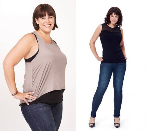
The basic method of treating obesity is conservative treatment consisting of diet, increased physical activity and pharmacotherapy, etc. Numerous empirical studies show that the effectiveness of this method ranges from 5-10% (long-term results of conservative treatment of obesity), which means that after 5 years only 5-10% of patients maintain the weight achieved during this type of treatment. Long-term results after bariatric surgery are 80% effective.
While conventional obesity therapy generates minimal weight loss for the patient, surgical treatment of obesity guarantees a weight loss of 40 to 87% depending on the type of surgery performed. It should also be noted that, in addition to directly affecting the weight of patients undergoing these procedures, surgical treatment of obesity also contributes to the resolution or significant improvement in the area of obesity co-morbidities, as well as reduces the mortality rate of obese patients.
Operations used in the surgical treatment of obesity, both classic and minimally invasive-laparoscopic, are divided according to their mechanism of action into:
Restrictive operations: they are performed only within the stomach. Significantly limit the patient's ability to eat food as a result of surgically dividing or resecting the stomach using a stretcher and / or metal staples.
- vertical gastric plastic surgery (Vertical Banded Gastroplasty - VBG)
- putting on the adjustable gastric band (AGB)
- Sleeve gastrectomy (SG)
Restrictive - disabling operations: they consist of producing a 20-30ml reservoir in the upper part of the stomach or a stomach cut of 4/5 (restrictive task) as well as enteric-intestinal anastomosis type Roux-en-Y with a specified length of alimony and enzymatic loop.
- gastrointestinal bypass (Roux-en-Y gastric bypass -RYGB)
- bile-pancreatic exclusion with bypass of the duodenum (biliopancreatic diversion - duodenal switch, BPD-DS)
- biliary-pancreatic exclusion consisting in cutting out 2/3 of the stomach and significantly shortening the gastrointestinal tract, which leads to the exclusion from the passage of a significant, specified length of the small intestine loop; also achieved results regarding weight loss such as regression of the components of the metabolic syndrome are very good

Other bariatric operations.
Other baritric operations are a group of procedures whose long-term results are not satisfactory. They should be treated as preparations for another bariatric procedure (intraglottic balloon) or treatments in the experimental phase (vagus nerve stimulators).
Selecting the operation method.
Each of these types of surgery can be used to treat obesity. After surgical treatment, assessing the percentage of excess weight loss (EWL), we achieve the greatest weight loss after performing biliary-pancreatic BPD-DS, and then bypassing gastrointestinal RYGB, vertical gastrectomy, and the smallest after putting on an adjustable band stomach (AGB) and sleeve gastrectomy (SR).
Bariatric surgery is a logical, reasonable, reproducible and safe method of treating obesity in patients whose attempts at conservative treatment have failed, and patients are convinced of the need for bariatric surgery.
Despite the enormous progress in bariatric surgery, the use of the latest surgical methods and a modern look at the problem of obesity, we must state that there is no golden surgery in the treatment of obesity, and both patients and the surgeon must decide on the choice of surgery treatment and inform the patient which of them would be best for a given patient.
Serdecznie zachęcamy do obejrzenia filmu z zabiegu Beskidzkiego Centrum Laparoskopowego z Centrum Leczenia Otyłości, który wykonał zespół dr Michała Dyaczyńskiego.
Reception doctors in the office
PhD in medical sciences
Michał Dyaczyński
specialist in general surgery
M.D.
Mirosław Kawulok
specialist in general surgery
